gallery
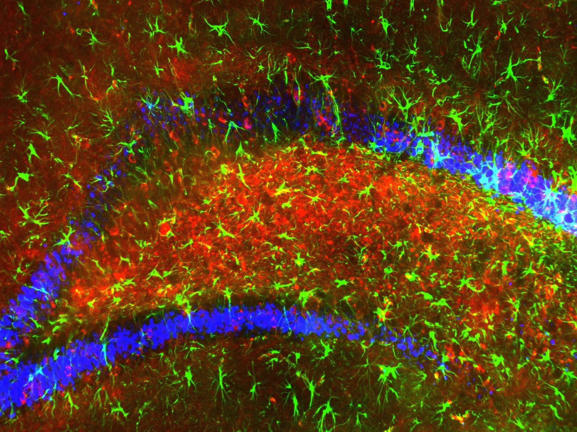
Fluorescent microscope image of neural tissue with green neurons, red glial cells, and blue background staining.
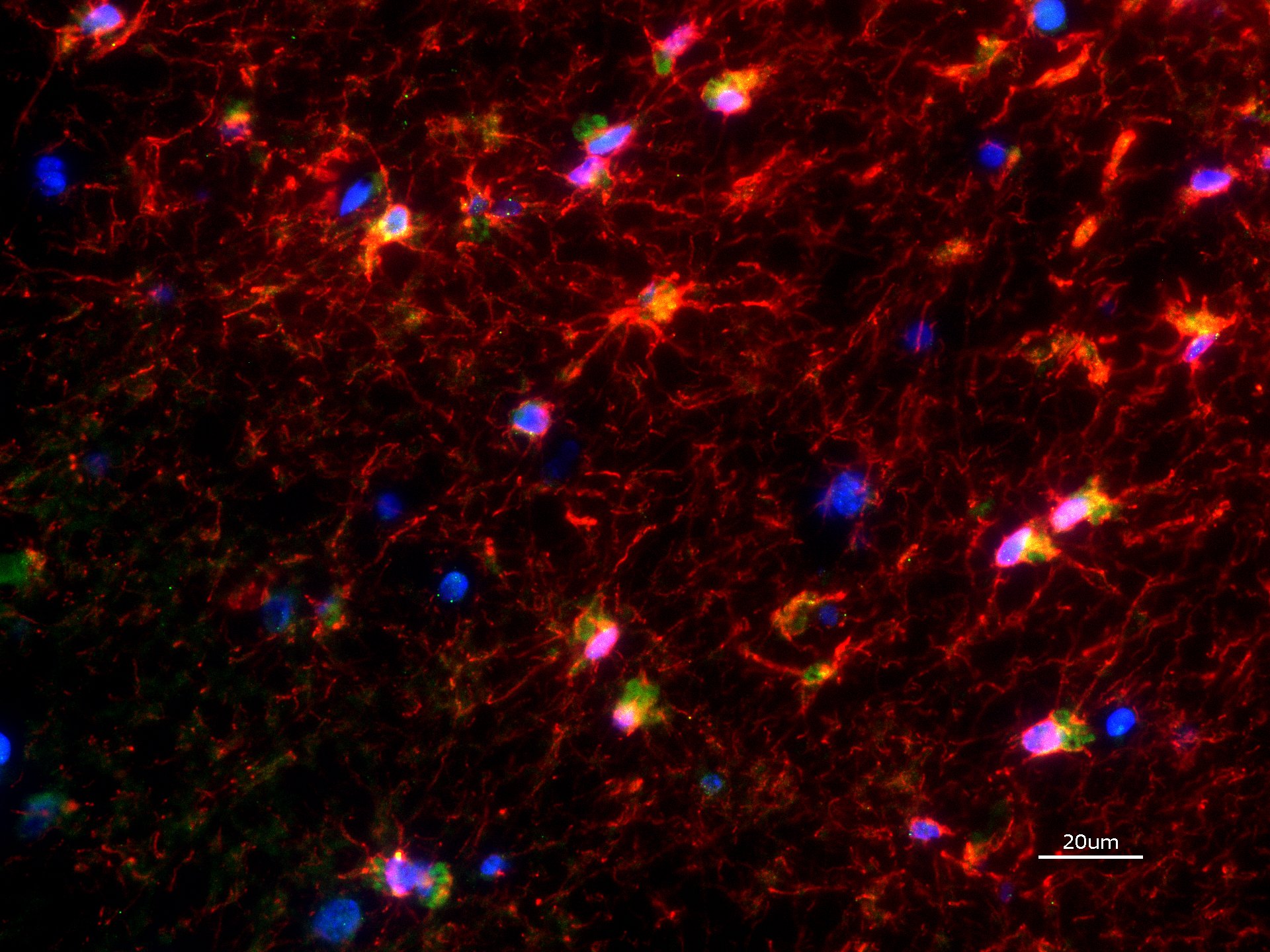
Microscopic view of neural cells with red, green, blue, and yellow fluorescent staining, showing a network of interconnected neurons with cell bodies and extensions.
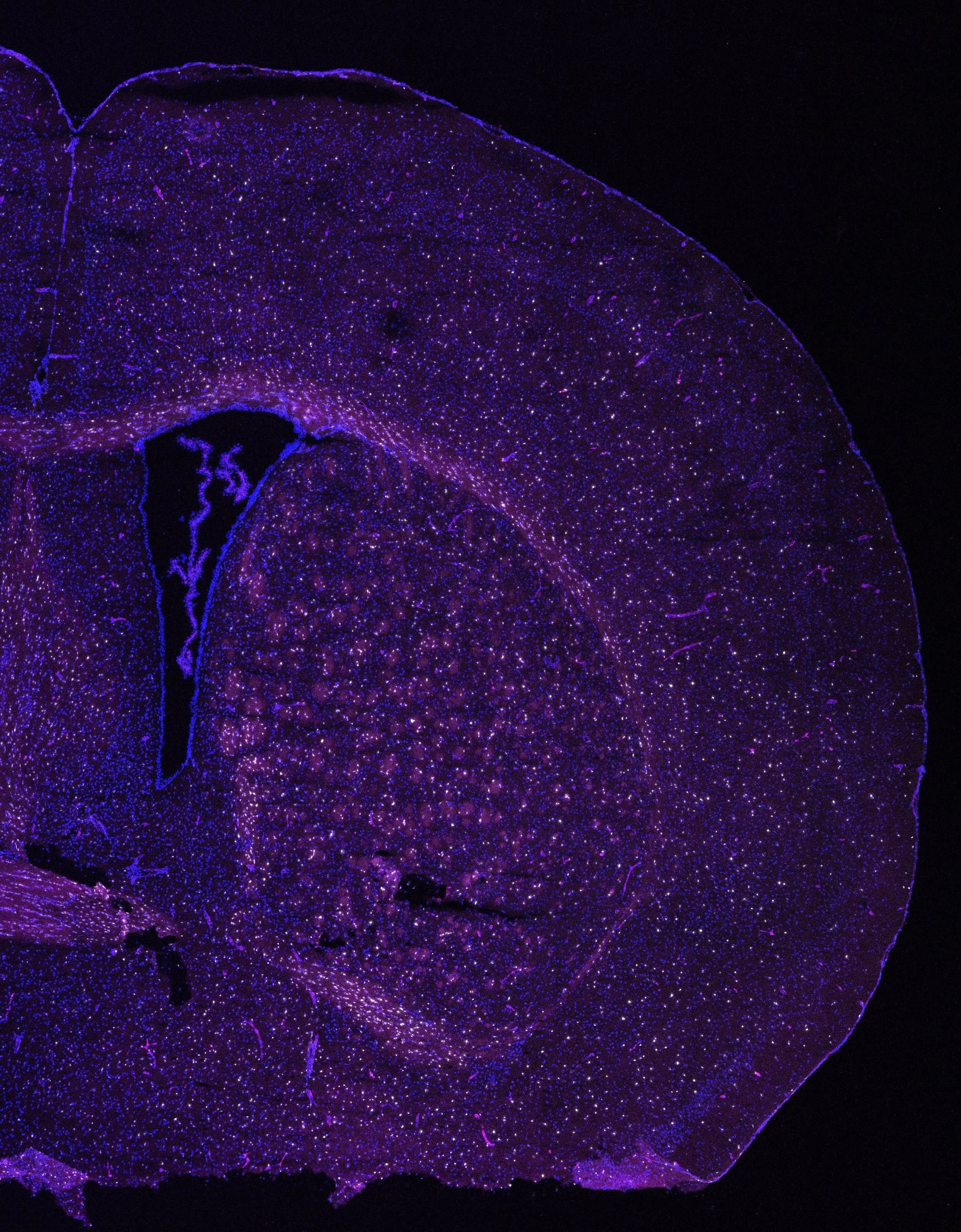
Close-up of a biological tissue sample stained with fluorescent dyes, showing purple and blue cellular structures against a black background.
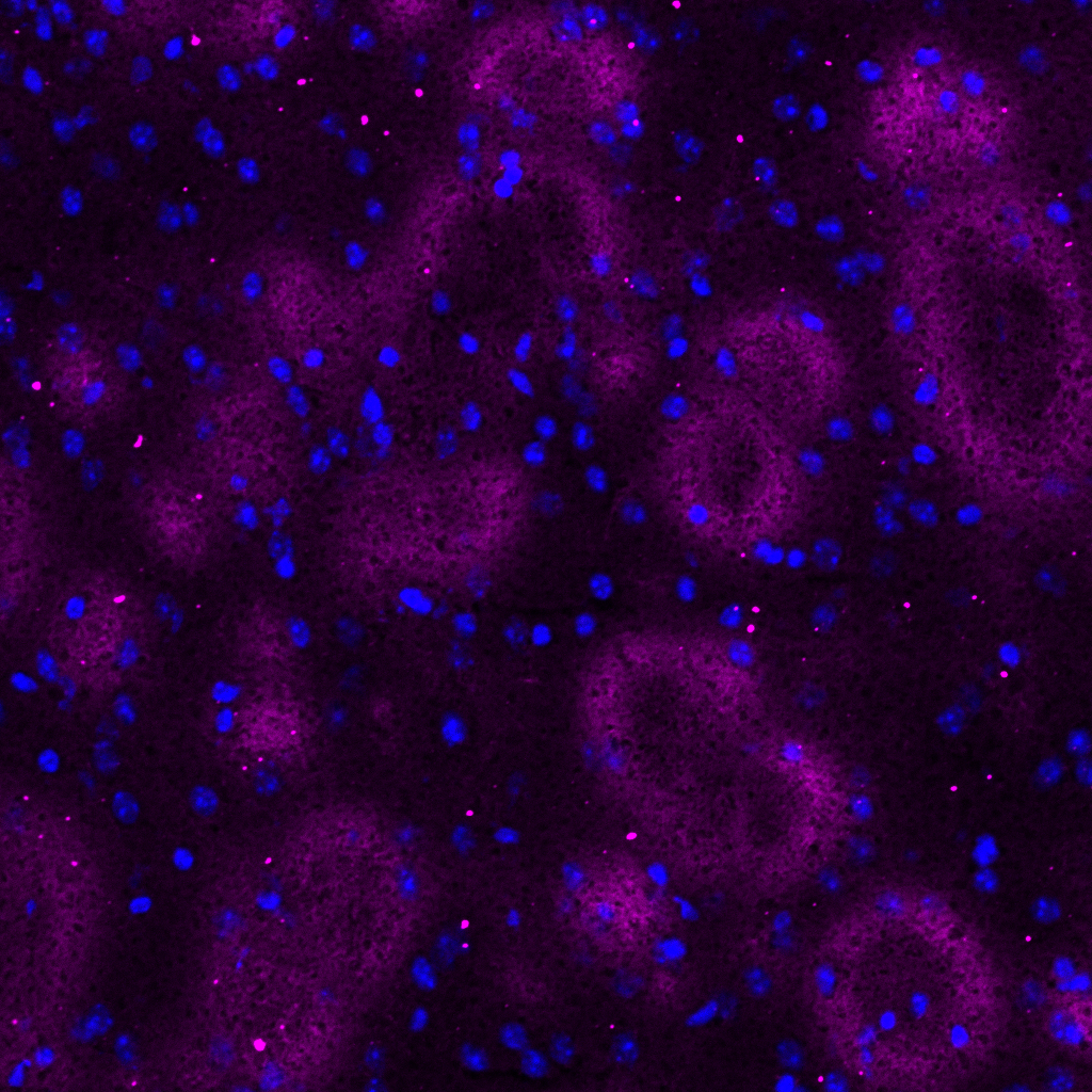
Close-up of a surface with purple and blue glowing spots, possibly a microscopic view of cells or bacteria

Fluorescent microscope image of a biological tissue sample showing various cell structures in green, purple, and blue colors, with a scale bar of 500 micrometers.